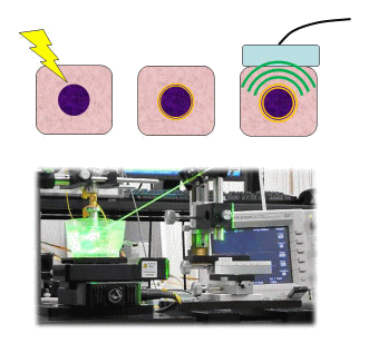
Photoacoustic Imaging
Selective and functional imaging technology for capillary network and erthrocytes
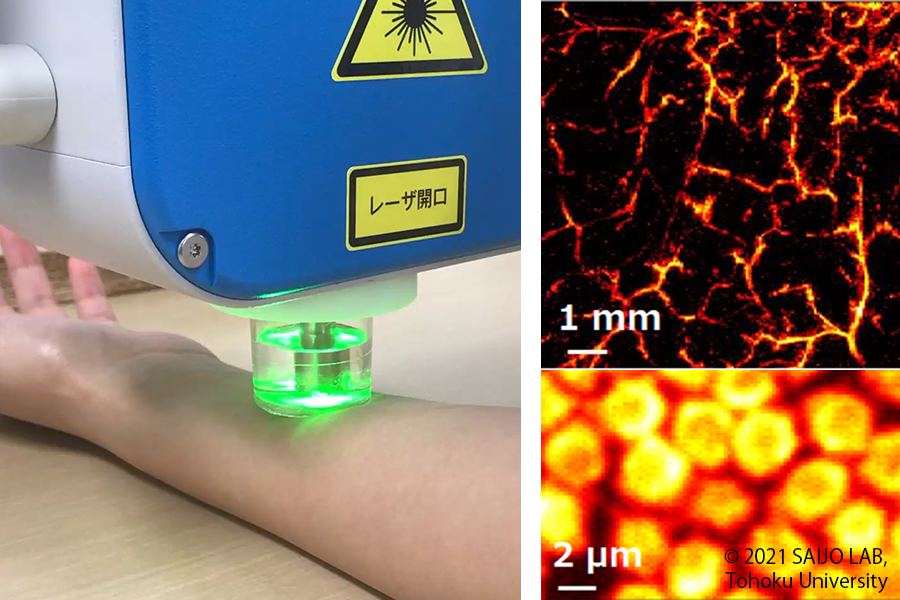
By irradicting the tissue with nanosecond pulse laser, the tissue momentarily undergoes thermal expansion and generates ultranonic waves. This phenomenon is called "Phtoacoustic effect", and the technology for capturing and imaging the generated ultrasonic waves with an ultrasonic sensor is called "Photoacoustic Imaging." Since the photacoustic effect depends on the absorbance of the object, it is possibble to image only blood vessels from inside tissue, for example, by using laser beam with a wavelength that blood absorbs well. In our laboratory, we have developed a variety of photoacoustic imaging systems, and have successfully images various scales from capillary networks to individual red blood cells. We are also working on functional imaging such as visualization of ixygen saturation distribution in vascular networks by using multiple laser beams.
Photoacoustic effect
By irradiating tissue with nanosecond pulses of laser causes instantaneous thermal expansion of the tissue, whihc generates ultrasound. Photoacoustic imaging is an imaging method that utilized this photacoustic effect. Usually, the spatial resolution of light is higher than that of ultrasound when performing tissue diagnosis, but light can only penetrate tissue up to about 2 mm, so depth is an issue. In photacoustic imaging, very small focus is possible because laser light is usesd on the way ther, and ultrasound is used on the way back, so attenuation in the tissue is small. As a result, imaging with twice the depth of field of regular laser light is possible. In addition, applying the differences in the spectra of oxidized and reduced hemoglobin, it is possible to analtze not only blood flow information but also tissue functions.