Medical imaging technology that can evaluate the condition of organ in the body without harming the body is indispensable to modern medicine. In particular, in-vivo imaging technology using ultrasound is playing an active role in various medical fields, from emergency medicine to home healthcare, as a real-time, safe, and highly portable means of examination. The saijo laboratory is challenging to expand the world of life phenomena visible by ultrasound while utilizing the advantages of existing ultrasound imaging.
More detailed spatio-temporal variablity of flow in the body, clearer images of capillary networks of tens of micron, clearer images of structural changes inside living cells, and instantaneous images of the three-dimensional structure of blood vesseels.
In order to realize the various requirements in medicine, We will create future medical imaging technology by integrating a wide range of engineering approaches, from ultrasound transdicer design, signal and image processing, optical systems, and artificial intelligence.
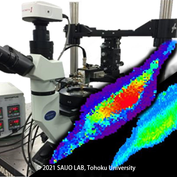
Acoustic Microscope
The spatial resolution of an ultrasound image increases in proportion to the freqeuncy of the ultraosund waves. Conventional frequencies used in medical ultrasound imaging range from 2 to 15 MHz, but in our laboratory, we use to 300 Mhz We are developing systems and signal processing technologies for "acoustic microscopes," which can visualize the microstructure of an object on a micron scale using a MHz ultrasonic transducer.
一Unlike ordinary optical microscope, ultrasound microscopes can visualize the three-dimensional distribution of mechanical properties such as hardness and viscoelasticity of tissue sections without staining, and are expected to be used in a wide range of healthcare fields such as tissue diagnosis of tumors, diagnosis of srterial plaque, and measurement of skin moisture content.
Ultrasound Signal Processing
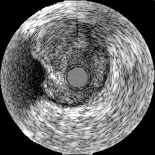
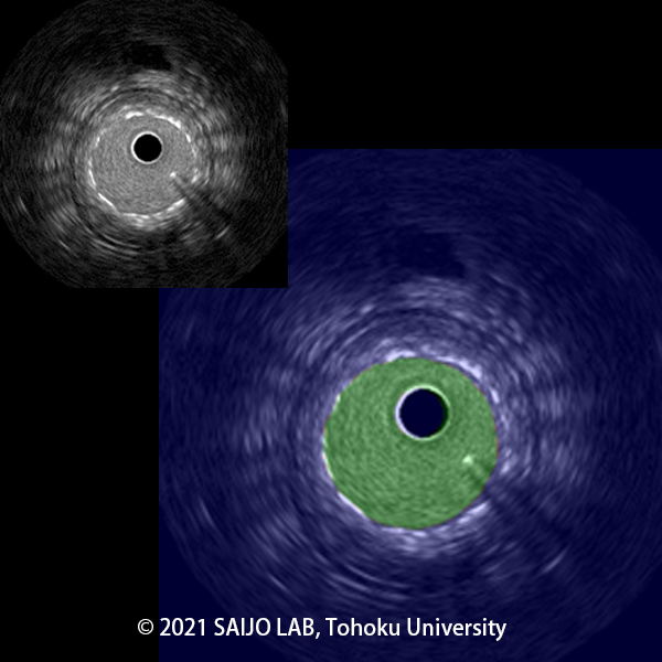
Ultrasound images typically show the intensity of ultrasound reflection in grayscale, displaying information about the shape and size of the tissue. Parameteric imaging is a method of diagnosing histological properties by imaging parameters such as frequency information and minute movements of blood vessles due to the load of blood pressure, as well as the strenth of ultrasonic reflection.
Applying this technique to intravascular ultrasound,parameters such as integrated backscatter (IB), tissue strain, virtual Histology, two-dimensional tissue velocity, and attenuation imaging enable automatic diagnosis of atherosclerotic tissue.
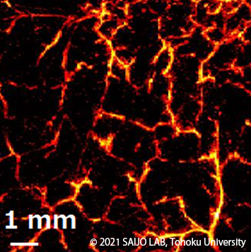
Photoacoustic Imaging
By irradicting the tissue with nanosecond pulse laser, the tissue momentarily undergoes thermal expansion and generates ultranonic waves. This phenomenon is called "Phtoacoustic effect", and the technology for capturing and imaging the generated ultrasonic waves with an ultrasonic sensor is called "Photoacoustic Imaging." Since the photacoustic effect depends on the absorbance of the object, it is possibble to image only blood vessels from inside tissue, for example, by using laser beam with a wavelength that blood absorbs well.
In our laboratory, we have developed a variety of photoacoustic imaging systems, and have successfully images various scales from capillary networks to individual red blood cells. We are also working on functional imaging such as visualization of ixygen saturation distribution in vascular networks by using multiple laser beams.
Blood Flow visualization in the body
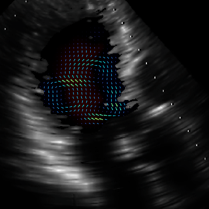
When an object moving inside the body, such as blood, is measured using ultrasound, the Doppler effect causes a frequency change in accordance with its speed. This change in frequency is visualized as a color Doppler image, whihc is one of the strengths of medical ultrasound imaging. By developing this technology, our laboray is conducting reserach on imaging the blood flow inside the heart, microvessels in the periphery, urine flow, and various other "flows" in the body more clearly and in detail.
For example, we have developed a technique called Echo-dynamograph (EDG) to visualize the vortex flow and jet flow inside the heart, and to use quantitative analysis of the flow for more detailed evaluation of cardiac motion.
Application of Artificial Intelligence AI in medicine
With the increasing importance of early diagnosis of diseases, daignostic support technologies for artificial intelligence (AI) such as deep learning continue to evolve day by day. In our Laboratory, we are working on optimizing AI models and learning methods that take into account the physical mechanism of image features in order to improve the reliability of diagnostic systems for ultrasound and X-ray images using AI technology. We are also aiming to create a mechanism that allows computers and people to collabrate efficiently by developing technology that visualizes the uncertainty of AI predictions results.
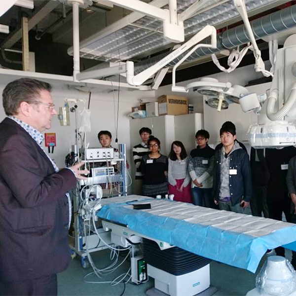
Medical Engineering Education and Social Contribution
As a part of the courses offered by the Graduate School of Biomedical Engineering, Tohoku University, we are creating a place where students of biomedical engineering and medical professional can lear from each other, think, and create new seeds of medical technology. We are also sending students from the Graduate School of Biomedical Engineering to a university in Netherlands, where biomedical engineering and entrepreneurship are flourishing, to promote academic exchange with local researchers and biomedical engineering students.
In addition, we provide human and technical support to various communities, such as holding information exchange meetings on ultrasound technology, mainly for cardiologists and clinical laboratoy technicians, and medical engineering education for working people.
